DeepTech deep technology MIT Technology Review exclusive partnership
 The "mouth" of a mosquito is our most common microneedle
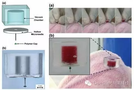
Editor's note: Scientists from the University of British Columbia (UBC) in Canada and the Paul Scherrer Institute (PSI) in Switzerland have invented a new medical microneedle system (see picture below) It can be used for painless subcutaneous testing and painless injection. Because its production cost is not high, it can greatly improve the patient's treatment experience, so it has a good chance to replace the existing traditional medical equipment and become the next generation of mainstream skin test and Injection system .
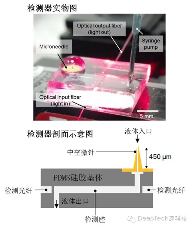
The newly invented medical microneedle system, the upper picture is the actual photo, the base material of the system is a kind of commonly used silica gel used as PDMS, and there is a cavity for liquid to flow through. The lower picture is a schematic cross-sectional view of the system. When used as a detector, the metal microneedle (with special treatment on the inner surface) penetrates the outermost stratum corneum of human skin, and draws a small amount of blood (less than 1nL) from the epidermis or dermis. When blood passes through the detection cavity, a beam of laser light enters the detection cavity through the optical fiber and then passes out, and enters the detection device from the other end of the optical fiber. The blood components can be analyzed by the absorption rate of the laser. If it is to be used for subcutaneous injection, laser equipment is not needed, and the drug can be directly introduced from the liquid outlet in the above picture, and then it can be injected into the patient through a microneedle. Image source: SA Ranamukhaarachchi, et al, Scientific Reoports 2016
The above-mentioned medical microneedle system related papers were published in "Scientific Reports" on July 6. The system successfully reduced vancomycin (vancomycin, an antibiotic, which may cause allergic reactions to some people. In severe cases, The blood required for the skin test is reduced by at least 50,000 times (50-100 μL is reduced to less than 1 nL), and the detection limit (lowest detectable concentration) has been increased by more than 13 times .
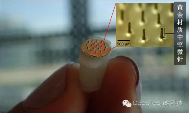
The miniature hollow needle is made of gold, which is convenient for adsorbing different biochemical reagents for medical testing. Image source: SA Ranamukhaarachchi, et al, Scientific Reoports 2016
The picture above shows one of the core components of the whole set of equipment-hollow micro-needles. These needles with a length of less than half a millimeter can break through the hardest stratum corneum in human skin, down to the capillaries of the epidermis and dermis. Minimize blood draw or administration, so as to achieve minimal harm to the human body .
The emergence of micro-needles saves patients with "injection phobia", but also greatly reduces the pain of some patients (such as diabetes) who require frequent subcutaneous injections. Since it was proposed at the beginning of this century, it is getting closer and closer to full-scale application now, and is about to become one of the mainstream medical methods. Those things about micro needles are worth talking about here.

In fact, everyone who has seen the disease knows that the hospital has two major difficulties: injections and medicine. The pain of taking medicine is too easy to solve, just add a sugar coating or a capsule. But if taking medicine can solve all problems, why do we need injections?
Oral medicine has many limitations. Before it acts on the human body, the metabolism of the human body and the decomposition and absorption of various enzymes such as saliva and intestine reduce the use of medicine. In addition, some oral drugs have a series of side effects such as irritation of the intestines and stomach and poisoning of the liver.
Therefore, in many cases, it is necessary to resort to less popular injections. Injection administration, especially intravenous injection, can deliver the medicine to the lesion with the highest efficiency. In addition, needles are the only sampling method for blood tests so far.
However, when the needle penetrates the skin, it is often accompanied by discomfort or even tingling. In the hospital, there are frequent cases of doctors or nurses being stabbed by needles due to careless operation. So, is there a way that is painless and can achieve efficient drug delivery?
There are methods, and they have a long history. This method is called Transdermal Drug Delivery. In words that everyone can understand, it is a plaster . The drug molecules in the applied plaster can diffuse into the capillaries under the surface skin and enter the blood circulation.
However, the biggest problem with these plasters is that the administration efficiency is not high . Human skin has a dense stratum corneum, and it is difficult for drug molecules with larger molecular weights to enter human capillaries through diffusion. Therefore, these plasters are often limited to the delivery of some drugs with relatively small molecular weights, and they are useless for some drugs or proteins with relatively large molecular weights.
To further solve the above problems, then only the protagonist of this article, Microneedle (Microneedle) came out.
As early as 2000, researchers at the Georgia Institute of Technology in the United States proposed that the use of needles with a length of about hundreds of microns (~400-800μm, or 0.4-0.8 mm) to pierce the skin can form a micron size in the stratum corneum of the skin. channels, thereby significantly improving the transdermal patch paste drug penetration, and to achieve include nucleic acids, antibodies, the DNA administration and the like insulin biomolecules.
What's more attractive is that these micro-needles are almost painless , and the skin trauma is much smaller than that of conventional hypodermic needles. It is a good news for patients with fainting needles, great!
Simply put, as a minimally invasive drug delivery method, microneedle drug delivery not only has the advantages of easy operation and painless transdermal drug delivery, but also has an effect comparable to conventional needle drug delivery.
My goodness, are micro needles really so amazing? Why can micro-needles do painless drug delivery, but not conventional needles?
This has to start with the structure of the skin. The human skin is a multi-layered tissue. On the outermost layer is the stratum corneum with a thickness of about 10-20 microns, which is the outermost and most important barrier of human skin. This layer is very dense and prevents the diffusion of most molecules into the skin. Below the stratum corneum is the upper epidermis, which is about 30 to 130 microns thick. Neither the stratum corneum nor the upper epidermis contains nerve endings and blood vessels. Below the upper epidermis is a dermal layer about 1 mm thick. The dermis contains capillaries and a small number of nerve endings.
As mentioned before, the length of the micro-needle needle is about 400~800 microns, so this layer is also the limit of the micro-needle skin puncture. Due to the small size of the micro-needle, the stimulation of the nerve endings in this layer is very limited. .
According to some researchers, when the micro-needle punctured the dermis, the subject felt a foreign body sensation, but did not feel pain. Below the dermis is the subcutaneous tissue. This part is rich in nerve endings and thick blood vessels, and this is the area where the needle is located during subcutaneous injection.
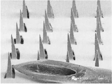
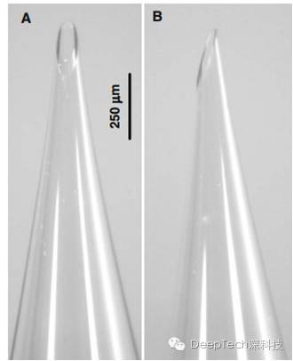
Due to the relatively large size of the needle for subcutaneous injection, it will inevitably stimulate the nerve endings and cause a tingling sensation. Below is a scanning electron micrograph comparing the needle used in conventional subcutaneous injection with the micro needle.
The thick hollow pyramid at the bottom of the picture is a conventional needle, and the relatively small structure on its side is a microneedle array. It can be said that the most essential reason for the completely different experience of conventional needles and micro needles is the huge difference in size.
Schematic diagram of the structure of human skin, from top to bottom are Stratum corneum, Living epidermis, Dermis and Hypodermis. Picture source: N. Roxhed, et al. J. MEMS, 2007
Comparison of scanning electron microscope photographs of solid microneedles and conventional needles, image source: W. Martano, et al, Pharmaceut. Res., 2004
Still find it hard to imagine the size of the microneedle? Still can’t understand why a needle that looks so sharp doesn’t hurt if it penetrates the skin?
In fact, there are many examples in nature. The most common one is probably the mosquitoes who have fallen in love with us for a lifetime . The mosquito can be regarded as a "flying needle", and its mouth needle for blood sucking is an excellent example of hollow microneedles. Its stylus is about 1~2 mm long and 50~100 microns in diameter.
The image below is a scanning electron microscope image of a mosquito needle. Most of the microneedles are about the same size as the mosquito's stylus, but the length is slightly shorter, about 300 to 700 microns.
Recall that when we were bitten by a mosquito, we would have some slight abnormality at most without obvious pain. (Note: The redness and swelling formed after a mosquito bite is a human inflammatory response to certain proteins in the mosquito’s saliva. The bite process is irrelevant).
The principle of the painless microneedles we are talking about is similar to that of mosquito bites.
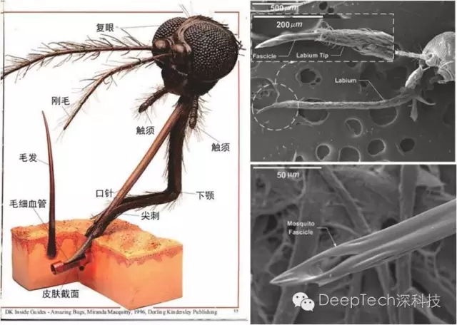
Left: schematic diagram of the mosquito’s anatomical structure, right: scanning electron microscope image of the mosquito’s stylus, image source: M. Ramasubramanian, et al. Bioinspir. Biomim., 2008
Returning to the topic of microneedles, after more than ten years of continuous research and development, the current microneedle family has developed four types: Solid Microneedle, Coated Microneedle, and Dissolved Microneedle (Dissolving Microneedle) and Hollow Microneedle, as shown in the diagram below.
Four schematic microneedles, from left to right are solid microneedles coated microneedles, the microneedles were dissolved with hollow microneedle Source: Y.Kim, et al Adv Drug Deliv Rev., 2012....
The application of micro-needles is no longer limited to skin puncture at the beginning of its creation. Their potential applications include vaccination, topical administration (such as mouth, eyes, etc.), and health monitoring.
Compared with the traditional injection method, the most prominent feature of the micro-needle is that it is a minimally invasive drug delivery method. The benefits of reducing the wound are obvious: it reduces the pain of the patient and shortens the wound recovery time . It is reported that the shielding effect of the stratum corneum is restored two hours after the use of the microneedles.
Compared with oral drugs and transdermal drug delivery, micro-needles have more ideal drug delivery effects. Therefore, from these two aspects, the front (money) scene (route) of microneedle is very bright. Next, the author will briefly introduce the characteristics of these four different needles.
Solid microneedle
The solid microneedle, as the name implies, is that the microneedle structure is solid. Speaking of it, the function of a solid microneedle is very simple. It is to pierce the skin and form micron-level channels on the skin surface, which can significantly improve the ability of the drug to diffuse into the skin.
With the continuous development of photolithography and micro-electromechanical systems, researchers have realized solid microneedles made of silicon, metal (such as stainless steel), and polymers.
The picture below is a picture of a solid microneedle jointly developed by researchers from the University of Kentucky and Georgia Institute of Technology. This microneedle patch contains 50 microneedles made of stainless steel. Each microneedle has a height of 620 microns, a bottom width of 160 microns, and a tip radius of less than 1 micron. The image on the right is the image after dyeing the skin after using the microneedle. It can be found that all 50 microneedles have pierced the skin.
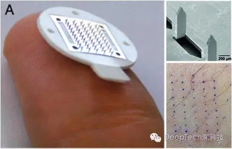
The image of the solid microneedle (left), the scanning electron microscope image of the solid microneedle (upper right), the image of skin staining after using the microneedle, and the blue dot is where the microneedle is inserted (lower right). Image source: D.Wermeling, et al. Proc. Natl. Acad. Sci, 2008
Coated microneedles
Since the solid microneedle still needs to be put on the place where it punctures the skin after use, it is not so convenient to use. So, is it possible to integrate these two steps? These thoughts gave birth to coated microneedles.
To put it simply, coating microneedles means to coat the drug on the solid microneedles. In this way, when the microneedles pierce the skin, the drug can freely diffuse inside the skin, and there is no need to apply plasters, which is convenient for the patient. At the same time, the use efficiency of microneedles is also improved.
Of course, when designing and preparing a coated microneedle, it is not as simple as directly coating the drug on the microneedle. How is the combination of drugs and microneedles? Will the two react? After piercing the skin, can the drug be separated from the microneedle and enter the blood? These many factors must be considered at the beginning of the design.
In most cases, ordinary solid microneedles must undergo appropriate surface modification treatment before they are used as coated microneedles.
Coated microneedle photos, where (A) is the scanning electron microscope image of the microneedles before the flu vaccine is coated, (B) is the macro photo, and (C) is the image after the drug is coated. Image source: Y.Kim, et al. J.Control. Release, 2010
Dissolving microneedles
The two types of microneedles mentioned above, solid microneedles and coated microneedles, will be contaminated with blood after use. To avoid the spread of diseases that may be caused by this, these microneedles will be discarded, just like those common needles. Like that. As a result, the use of microneedles will inevitably bring harmful medical waste.
So, is there a better solution? The researchers proposed that biodegradable materials can be used to make microneedles, which is the origin of the dissolved microneedles.
The structure of this microneedle is similar to the coated microneedle mentioned earlier. The main difference is that this type of microneedle is due to harmless degradable polymer materials (such as maltose polymer, polylactic acid, polyhydroxy acid, etc.) Made.
Because most of these degradable polymer materials are soluble in water. Therefore, after entering the blood environment in the human body, these needles will decompose and release drugs without producing harmful biological waste.
Dissolving microneedles made of maltose polymers. Image source: K. Lee, et al. Biomaterials, 2012
Hollow microneedle
The aforementioned microneedles can essentially be regarded as solid microneedles and their derivatives. Although these microneedles can promote and enhance the efficiency of transdermal drug delivery, the disadvantage is that the delivery of drugs depends on passive diffusion and cannot achieve active control of the drug delivery rate like ordinary needles. On the other hand, these microneedles also The blood cannot be drawn. This type of microneedle was mentioned at the beginning of this article.
As we all know, human blood is an important medium in the human body and contains a lot of biological information that can be used for disease diagnosis and health monitoring.
People need a new device that can draw blood, but is not as painful as a traditional needle. In this context, hollow microneedles came into being.
Compared with solid microneedles, the biggest feature of hollow microneedles is that they have channels for liquid transmission. This feature enables the hollow microneedle to be fused with some miniaturized biological monitoring equipment to achieve continuous and real-time human health monitoring.
At the same time, by connecting the hollow microneedle with the drug storage capsule, it is also possible to realize the autonomous regulation of the drug delivery rate, which can achieve multiple benefits in one fell swoop. From the aspect of drug preparation, the hollow microneedle can directly adopt the existing subcutaneous injection drug formulation, which undoubtedly simplifies the use of the microneedle.
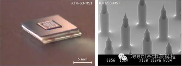
Scanning electron microscope images of the hollow microneedle patch (left) and the hollow microneedle array. Image source: N. Roxhed, et al. IEEE. Trans. Bio-med. Eng., 2008
After decades of development, micro-needles have begun to make their mark in health care and other fields. So let’s talk about what applications are there for micro needles?
Therapeutic drug delivery
For diabetics, insulin injection is undoubtedly a very important part of daily life. Compared with conventional needle subcutaneous injection, microneedle-based drug delivery is obviously a painless, minimally invasive and safer drug delivery method.
In the early days of microneedle research, many researchers have begun to pay attention to the direction of microneedle assisted insulin delivery.
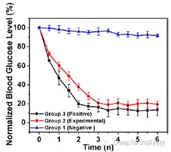
Researchers at the National University of Singapore conducted continuous observation and measurement of blood glucose levels in mice for 6 hours, and obtained the measurement results of blood glucose concentration as shown in the figure below.
Three groups of mice with diabetes symptoms were used in this experiment. The abscissa in the graph is time, and the ordinate is the blood glucose concentration. The blue curve is the negative control group (only insulin solution is dripped on the skin), the red curve is the experimental group (after using the microneedle, insulin solution is dripped on the skin), and the black curve is the positive control group (conventional needle injection) . It can be found that the blood glucose concentration of the experimental group is equivalent to that of the positive control group, which is significantly better than that of the negative control group.
In addition to insulin, solid microneedle pretreatment has a very significant effect on the transdermal delivery of many other therapeutic drugs, such as morphine antagonist naltrexone NTX, nucleic acid, lidocaine, influenza vaccine, etc.
Based on the experimental results of microneedle administration. Image source: Z. Xiang, et al. J. Micromech. Microeng., 2015
Health monitoring
As mentioned before, the human blood contains a lot of biological information that can be used for disease diagnosis and treatment and health monitoring. Speaking of blood tests, everyone is familiar with it. When I arrived at the hospital, I rolled up my sleeves, and the nurse sister looked for blood vessels and took out the needle to prick her skin. This sour feeling is really unforgettable.
The good news is that with the continuous development of bio-microelectromechanical systems, in the near future, hollow microneedles will be able to integrate with miniaturized biochemical testing equipment to achieve painless, real-time continuous health monitoring.
Recently, researchers have developed a handheld blood collection device using hollow microneedles.
Hand-held blood sampling device based on hollow microneedles. Image source: C.Li, et al., Lab. Chip., 2015
Local administration
Since the volume of the microneedle is much smaller than that of a conventional needle, it can achieve more precise local drug delivery. What attracted my attention the most was the administration to the eyes.
As we all know, due to the many fine and complex structures in the human eye and the shielding effect of the cornea, ocular drug delivery, especially the posterior chamber (including sclera, vitreous, etc.) has always been a very big challenge. The lesions of this part of the tissue caused by the et al. are an important factor in blindness. Our commonly used eye ointments and eye drops are often unsatisfactory for the actual administration effect of this part.
Used for hollow microneedles used in ocular drug delivery experiments. Image source: J. Jiang, et al., Pharmaceut. Res., 2009
Researchers from Georgia Tech and Emory University used hollow microneedles to achieve the goal of delivering drugs to the iris of the eye.
The results of the study confirm that the use of hollow microneedles can indeed deliver nano- and micro-scale particles to the iris of the eye.
Of course, the ocular drug delivery experiment is currently carried out in vitro, and there is still a long way to go before the real commercialization, that is, in vivo drug delivery. .